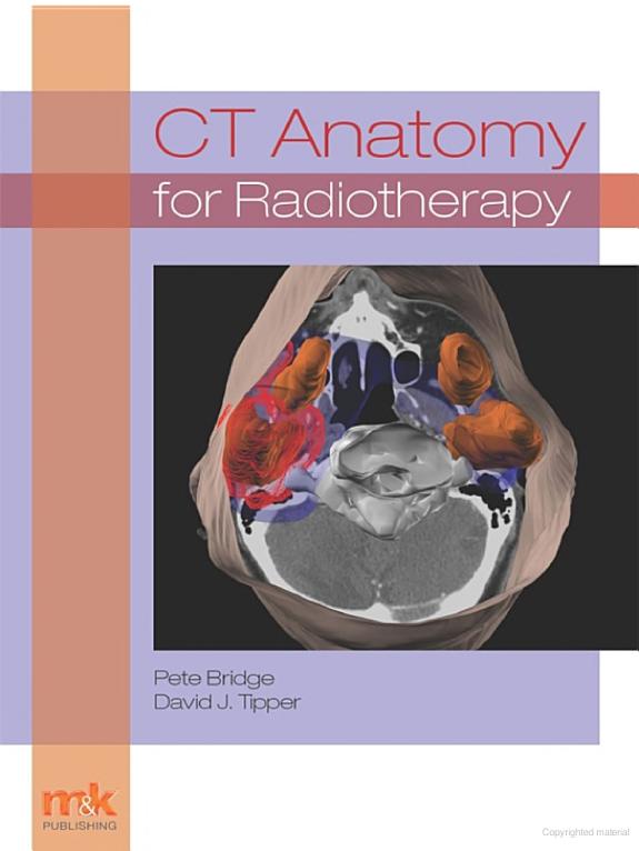
CT Anatomy for Radiotherapy
by Pete Bridge
302 pages· 2011· ISBN 9781907830549
About
Knowledge of CT anatomy is increasingly vital in daily radiotherapy practice, especially with more widespread use of cross-sectional image-guided radiotherapy (IGRT) techniques. Existing CT anatomy texts are predominantly written for the diagnostic practitioner and do not always address the radiotherapy issues while emphasising structures that are not common to radiotherapy practice. OC CT Anatomy for RadiotherapyOCO is a new radiotherapy-specific text that is intended to prepare the reader for CT interpretation for both IGRT and treatment planning. It is suitable for undergraduate students, qualified therapy radiographers, dosimetrists and may be of interest to Knowledge of CT anatomy is increasingly vital in daily radiotherapy practice, especially with more widespread use of cross-sectional image-guided radiotherapy (IGRT) techniques. Existing CT anatomy texts are predominantly written for the diagnostic practitioner and do not always address the radiotherapy issues while emphasising structures that are not common to radiotherapy practice. OC CT Anatomy for RadiotherapyOCO is a new radiotherapy-specific text that is intended to prepare the reader for CT interpretation for both IGRT and treatment planning. It is suitable for undergraduate students, qualified therapy radiographers, dosimetrists and may be of interest to oncologists and registrars engaged in treatment planning. All essential structures relevant to radiotherapy are described and depicted on 3D images generated from radiotherapy planning systems. System-based labelled CT images taken in relevant imaging planes and patient positions build up understanding of relational anatomy and CT interpretation. Images are accompanied by comprehensive commentary to aid with interpretation. This simplified approach is used to empower the reader to rapidly gain image interpretation skills. The book pays special attention to lymph node identification as well as featuring a unique section on Head and Neck Deep Spaces to help understanding of common pathways of tumour spread. Fully labelled CT images using radiotherapy-specific views and positioning are complemented where relevant by MR and fusion images. A brief introduction to image interpretation using IGRT devices is also covered. The focus of the book is on radiotherapy and some images of common tumour pathologies are utilised to illustrate some relevant abnormal anatomy. Short self-test questions help to keep the reader engaged throughout."
Discuss CT Anatomy for Radiotherapy with other readers
Join or start a book club for CT Anatomy for Radiotherapy on Readfeed. Live chat, shared reading progress, and AI discussion questions — free to get started.
Frequently asked questions
How do I join a book club for CT Anatomy for Radiotherapy?
Sign up free on Readfeed, then browse public clubs or start your own club with CT Anatomy for Radiotherapy as the current read. Invite friends with a share link and discuss together with live chat and AI discussion questions.
Can I discuss CT Anatomy for Radiotherapy with other readers online?
Yes. Readfeed book clubs let you chat live, share progress, and join discussions about CT Anatomy for Radiotherapy with readers worldwide — whether your club is virtual, in-person, or hybrid.
Is Readfeed free?
Yes. Creating an account and joining book clubs is free. Sign up to find readers who love the same books and start discussing today.